What Is a Slit Lamp? History, Types, and Clinical Uses
If you have ever had a comprehensive eye exam, you have likely sat behind a slit lamp — the instrument with two eyepieces that the doctor uses to look deep into your eye while a thin beam of light sweeps across the cornea, iris, and lens. But what exactly is a slit lamp, how does it differ from other ophthalmic instruments, and why is it considered the workhorse of every modern eye clinic?
This guide explains what a slit lamp is, traces its 100+ year history, breaks down the major types and components, and walks through the clinical examinations that depend on it. Whether you are a student entering optometry school, a clinic manager evaluating instruments, or a curious patient wanting to understand the equipment aimed at your eye, this article gives you a complete foundation.
1. What Is a Slit Lamp?
A slit lamp is a binocular biomicroscope specifically designed for examining the anterior segment of the eye — the cornea, iris, lens, and anterior chamber — under high magnification and controlled illumination. The name comes from its defining feature: the illumination system can project a narrow “slit” of light onto the eye, allowing the clinician to optically section the cornea and lens in cross-section, much like slicing a loaf of bread to see the interior.
A modern slit lamp combines three integrated systems:
- Illumination system — a halogen or LED light source with adjustable slit width, height, angle, and filters
- Microscope system — a binocular stereoscopic microscope with 5x to 40x magnification, usually with a continuous zoom
- Mechanical stage — a chinrest, forehead bar, and joystick-controlled base that position the patient and align the optics
2. A Brief History of the Slit Lamp
The slit lamp is older than most eye care professionals realize. Its development spans more than a century and tracks closely with the broader history of ophthalmology.
| Year | Milestone | Significance |
|---|---|---|
| 1911 | Allvar Gullstrand invents the slit illumination principle | First optical sectioning of the cornea; won Nobel Prize for ophthalmic optics |
| 1916 | Henker and Gullstrand build the first commercial slit lamp | Combined Gullstrand’s illuminator with a Zeiss binocular microscope |
| 1933 | Goldmann introduces the Haag-Streit 900 style slit lamp | Established the modern joystick-controlled mechanical stage still used today |
| 1980s | LED illumination and continuous zoom optics emerge | Longer bulb life, cooler operation, smoother magnification transitions |
| 2000s | Digital imaging integration becomes standard | Built-in cameras, beam splitters, DICOM output for EHR |
| 2010s+ | Portable and handheld slit lamps mature | Battery-powered units bring quality exams to pediatrics, bedside, and outreach |
The fundamental optical principle has not changed since 1911 — what has changed is ergonomics, illumination quality, and integration with digital diagnostics.
3. How a Slit Lamp Works
A slit lamp performs two simultaneous functions: it lights the eye with a precisely shaped beam, and it magnifies the resulting image through a stereoscopic microscope. The clinician’s ability to detect disease depends on mastering both.
Illumination techniques — the foundation of slit lamp diagnostics:
| Technique | How It Is Done | What It Reveals |
|---|---|---|
| Diffuse Illumination | Wide beam, low magnification, scattered light | Overall anterior segment overview; lid margins; conjunctiva |
| Optical Sectioning | Very narrow slit beam at 45° angle | Cross-section of cornea and lens; depth of opacities and scars |
| Sclerotic Scatter | Beam aimed at limbus (corneoscleral junction) | Subtle corneal edema, infiltrates, and microcysts |
| Specular Reflection | Angle set to reflect off endothelial layer | Corneal endothelial cell morphology; guttata in Fuchs dystrophy |
| Cobalt Blue + Fluorescein | Blue filter + yellow fluorescein dye | Corneal abrasions, ulcer borders, contact lens fitting, applanation tonometry |
A single comprehensive exam typically uses 4-5 of these techniques in sequence. Mastery of slit lamp technique is one of the first clinical skills optometry and ophthalmology students must develop.
4. Types of Slit Lamps
Not every slit lamp is the bulky chrome unit you see in every exam lane. The instrument comes in several form factors, each optimized for a clinical setting.
- Table-mounted slit lamps are the standard. They include a heavy base, binocular microscope, illumination arm, and patient chinrest assembly. Most have 5-step or continuous zoom magnification from 6x to 40x, halogen or LED illumination, and a joystick-controlled mechanical stage for fine positioning.
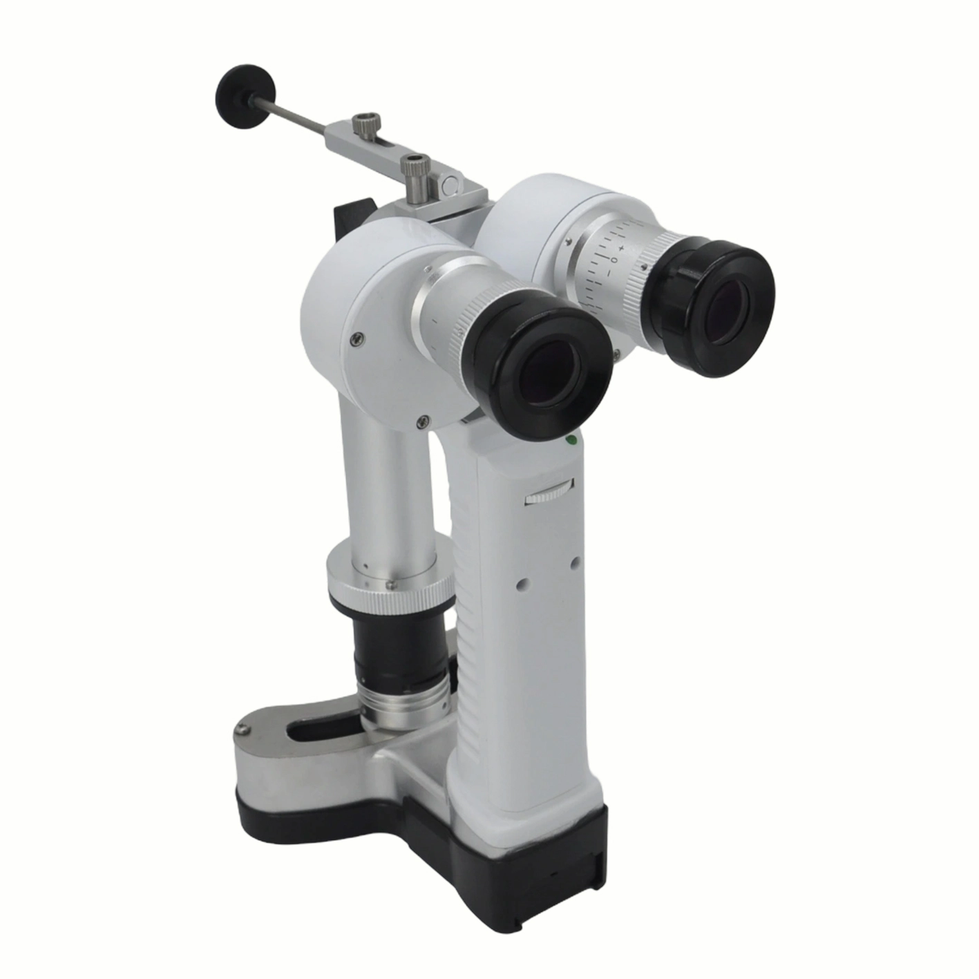
- Portable slit lamps use the same optical principles but shrink the form factor into a handheld unit with rechargeable battery, smaller magnification range (typically 5x to 10x), and integrated LED illumination. They trade some resolution for mobility — ideal for pediatrics, bedside exams, nursing home visits, and field clinic deployments where a table-mounted unit is impractical.
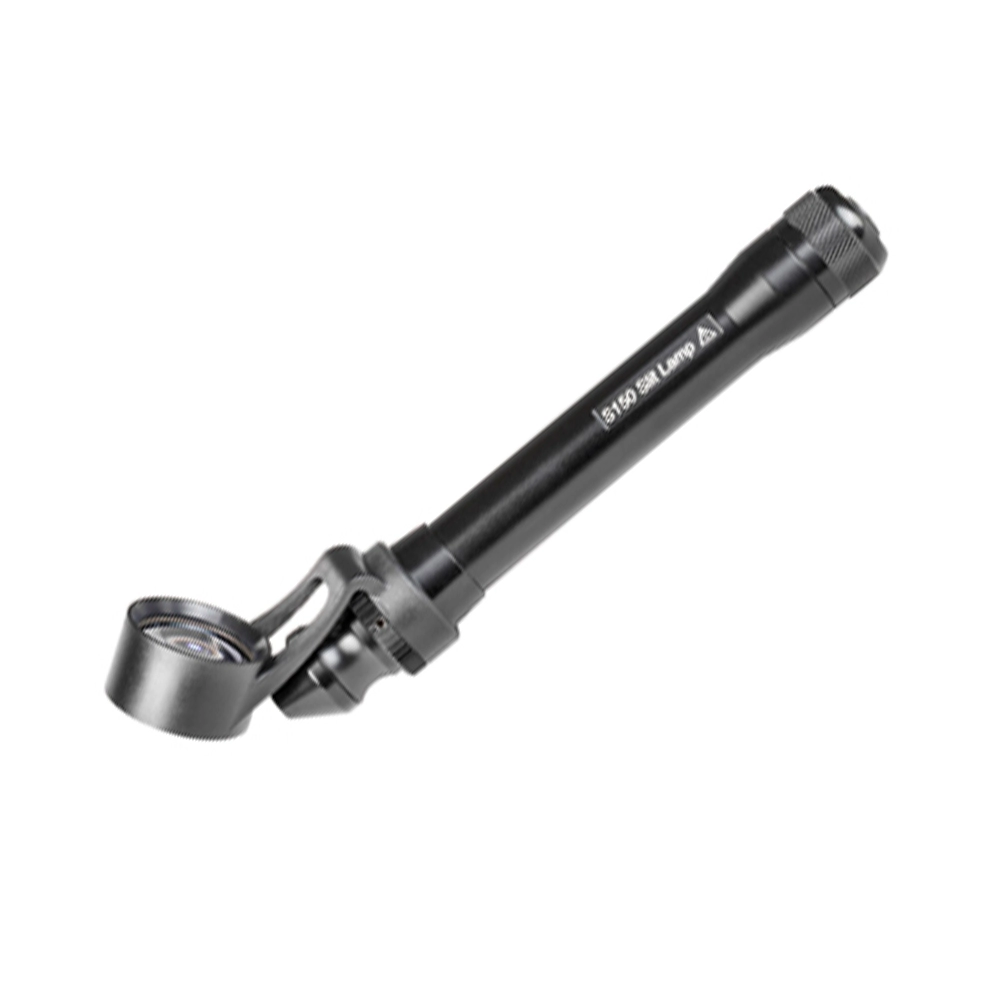
- Handheld pocket slit lamps are the smallest tier — pocket-size diagnostic instruments with 2x to 8x magnification, often with a single fixed slit width. They are intended for quick screenings and triage rather than comprehensive exams.
- Digital slit lamps replace one or both eyepieces with a high-resolution camera and display, allowing the examiner to view the eye on a screen, capture still images and video, and transmit findings to an EHR. Many digital units also support auto-focus and image-enhancement algorithms.
- Surgical slit lamps are specialized for procedures such as YAG laser capsulotomy, argon laser trabeculoplasty, and SLT. They add laser delivery optics, surgeon-friendly foot pedal controls, and enhanced safety filters.
5. Parts of a Slit Lamp
A modern table-mounted slit lamp has six functional component groups. Understanding what each does helps with operation, troubleshooting, and procurement decisions.
| Component | Function | Typical Specs |
|---|---|---|
| Illumination Arm | Holds the light source, slit mechanism, and filters; adjustable angle 0-90° | Halogen 6V/10W or LED 3-5W; slit width 0-9mm |
| Microscope Head | Binocular stereoscopic optics with continuous zoom or 5-step turret | 5x to 40x magnification; 12.5x or 16x eyepieces |
| Patient Assembly | Chinrest, forehead bar, fixation light; aligns patient’s visual axis | Adjustable height 60-90cm; black cup for ambient eye isolation |
| Mechanical Stage | Joystick-controlled base for X-Y-Z fine positioning | Travel range ~80mm X-Y; fine focus scale 0.1mm |
| Filters & Diaphragms | Cobalt blue, green (red-free), heat-absorbing, neutral density | 4-6 filter positions typical; interchangeable diaphragms |
| Imaging Port (Optional) | Beam splitter + camera mount for digital capture | C-mount or T-mount; 50/50 or 70/30 light split |
For digital workflows, the imaging port is the upgrade path: a beam splitter attaches between the microscope head and eyepieces, routing 30-50% of light to a DSLR, mirrorless, or industrial camera. This converts a traditional slit lamp into a documentation-ready clinical instrument.
6. What a Slit Lamp Is Used For
The slit lamp is the most frequently used diagnostic instrument in ophthalmology and optometry. Its clinical applications span from routine vision exams to specialized diagnostics:
- Comprehensive anterior segment examination — cornea, conjunctiva, iris, lens, and anterior chamber assessment at every routine eye exam
- Contact lens fitting — evaluating lens centration, movement, and fluorescein patterns (with cobalt blue illumination)
- Corneal disease diagnosis — detecting keratoconus, Fuchs dystrophy, dry eye syndrome, pterygium, and corneal ulcers
- Cataract grading — classifying nuclear, cortical, and posterior subcapsular cataracts per LOCS III standards
- Pre- and post-operative assessment — evaluating patients before refractive surgery, cataract extraction, corneal cross-linking, and glaucoma procedures
- Tonometry (with Goldmann applanation prism) — measuring intraocular pressure via the slit lamp mounted applanation tonometer
- Gonioscopy (with goniolens) — visualizing the anterior chamber angle to classify open-angle vs angle-closure glaucoma
- Fundus examination (with auxiliary 78D/90D lenses) — extending the slit lamp’s reach to the retina, optic nerve, and macula without a separate direct ophthalmoscope
7. How to Choose the Right Slit Lamp
Selecting a slit lamp depends on three factors: practice setting, patient volume, and integration needs.
| Use Case | Recommended Type | Key Features to Prioritize |
|---|---|---|
| Primary Exam Lane | Table-mounted, LED, continuous zoom | Reliable daily-use instrument; 10-20 year service life |
| Pediatrics / Bedside | Portable handheld | Rechargeable battery, fast warm-up, rugged case |
| Mission / Field Clinic | Pocket slit lamp + portable backup | Lightweight, battery-independent operation, dust resistant |
| Teaching Institution | Table-mounted + beam splitter + camera | Live video output for student observation; image archive for case reviews |
| Surgical Practice | Surgical slit lamp (YAG/SLT-ready) | Laser delivery optics, foot pedal controls, safety filters |
| EHR-Integrated Practice | Digital slit lamp with DICOM output | Direct image transfer to patient record; auto-naming conventions |
For most established optometry practices, a single high-quality table-mounted slit lamp plus a portable slit lamp for non-routine cases forms an effective instrument portfolio.
8. Frequently Asked Questions
What is the difference between a slit lamp and an ophthalmoscope?
A slit lamp is a binocular biomicroscope that examines the anterior segment (cornea, iris, lens) at high magnification under controlled illumination. An ophthalmoscope is a handheld monocular instrument that examines the posterior segment (retina, optic nerve, macula) through the pupil. The slit lamp can also view the posterior segment when paired with a 78D or 90D auxiliary lens, but the ophthalmoscope remains the traditional tool for direct fundus examination.
Does a slit lamp exam hurt?
No. A slit lamp examination is non-contact and painless. The patient simply rests their chin and forehead against the instrument while the clinician examines the eye with light. The only mildly uncomfortable moments are when dilating drops are applied before the exam (slight stinging for 2-3 seconds) or when fluorescein dye is used (temporary mild foreign body sensation).
Why is it called a slit lamp?
The instrument is named for its defining optical feature: the illumination system can project a thin vertical “slit” of light (0-9mm wide, adjustable) onto the eye. This slit allows the clinician to optically section the cornea and lens in cross-section, revealing the depth, location, and morphology of any pathology inside the transparent tissues.
Can a slit lamp detect glaucoma?
Yes, but only as part of a comprehensive exam. The slit lamp is used to assess anterior chamber depth (a risk factor for angle-closure glaucoma), perform gonioscopy to visualize the drainage angle, measure intraocular pressure via Goldmann applanation tonometry, and — with auxiliary lenses — evaluate the optic nerve head for glaucomatous cupping. Full glaucoma diagnosis requires all of these findings combined with visual field testing and OCT imaging.
How long does a slit lamp last?
A well-maintained slit lamp from a reputable manufacturer lasts 15-25 years in routine clinical use. The optics rarely fail; most replacements are driven by illumination technology upgrades (halogen to LED), digital integration needs, or wear on the mechanical stage from daily patient flow. Annual preventive maintenance costs are typically 1-3% of the instrument’s original purchase price.
How much does a slit lamp cost?
Slit lamp prices range widely based on type, brand, and features. Entry-level table-mounted units start around $3,000; precision mid-range models from Topcon, Zeiss, or Haag-Streit run $8,000 to $15,000; surgical-ready units with laser delivery reach $25,000+. Portable and handheld slit lamps range from $500 to $4,000 depending on magnification and illumination quality. For a detailed pricing breakdown across tiers and use cases, our companion guide covers the cost factors that determine slit lamp price.
Looking to add a slit lamp to your practice or replace an aging unit? Our team can help you compare options, evaluate workflow fit, and plan a procurement timeline. Contact us for a personalized recommendation.