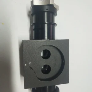
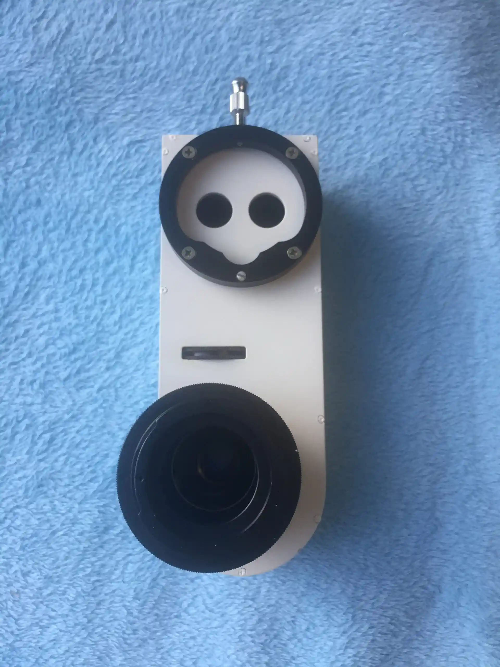
Integrated Camera Photography Beam splitter
An integrated beam splitter with built-in optical converter lens for high-resolution digital image acquisition during ophthalmic microscopy, featuring German-imported multi-coated lenses and plug-and-play installation.
Description
The Integrated Camera Photography Beam Splitter (YTH01) combines a beam splitter with an optical converter lens for high-resolution digital image acquisition during ophthalmic microscopy. Supplied by BrightVision Tech, a specialized ophthalmic equipment supplier, all lenses are imported from Germany with multi-coated, mildew-proof, and anti-reflection treatment for clear optical observation.
The unit features fine focus control and a compact, lightweight body. Its plug-and-play design requires no professional technician for installation — the optical alignment is pre-adjusted at the factory. The YTH01 is compatible with standard C-mount camera systems, allowing clinics to use their preferred camera body for image capture and documentation.
Key Features
- German-Imported Optics: Multi-coated, mildew-proof, and anti-reflection lenses sourced from Germany for color-accurate image capture
- Built-In Optical Converter: Integrated lens for direct high-resolution digital image acquisition without external adapters
- Plug-and-Play Installation: Pre-adjusted optical alignment — attach to the microscope and start capturing with no technician required
- Compact and Lightweight: Minimal footprint preserves microscope balance and maneuverability
- Fine Focus Control: Precise adjustment ring for accurate image framing and calibration
Specifications
| Parameter | Detail |
|---|---|
| Model | YTH01 |
| Optical Converter | Built-in |
| Lens Quality | German-imported, multi-coated |
| Camera Mount | Standard C-mount compatible |
| Focus Control | Fine focus adjustment |
| Installation | Plug-and-play, pre-aligned |
Applications
- Eye clinics — slit lamp photography for patient records
- Surgical microscopy — real-time image capture during ophthalmic procedures
- Clinical teaching — live image display for training and education
- Hospital ophthalmology departments — documentation of anterior segment findings
- Equipment distributors — imaging accessory for microscope product lines